Animal Cell Structures and Functions

Animal cell coloring key worksheet – Alright, let’s dive into the amazing world of animal cells – it’s like a microscopic city bustling with activity! Think of it as a super-powered, miniaturized version of a bustling metropolis, each component playing a vital role in keeping the whole thing running smoothly. We’ll explore the key players and their functions, comparing them to a plant cell along the way.
Animal Cell Organelles: Structure and Function, Animal cell coloring key worksheet
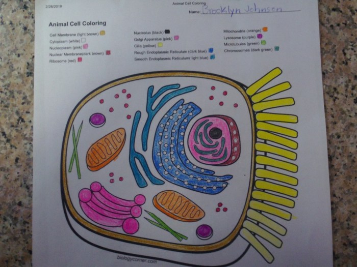
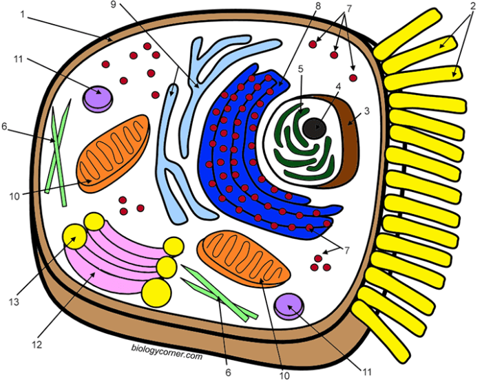
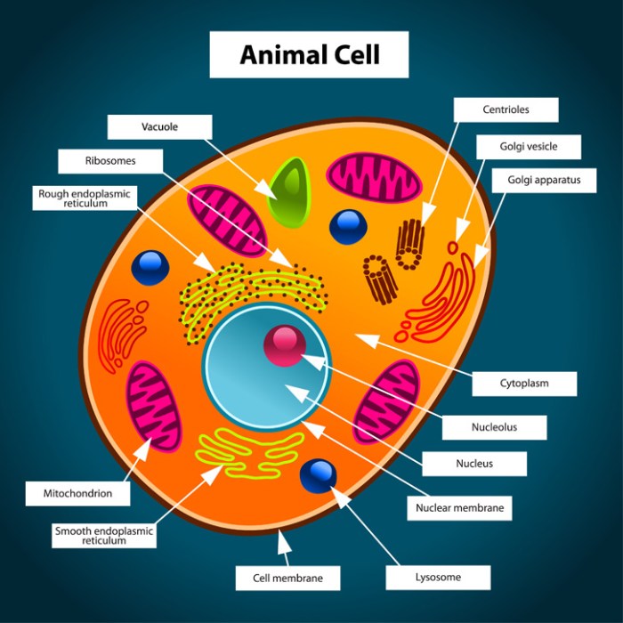
The animal cell is packed with organelles, each with a specific job. It’s like a well-oiled machine, where every part is essential. Let’s break down the key players: the nucleus is the control center, the mitochondria are the powerhouses, and the cell membrane is the bouncer, carefully controlling what enters and exits. The other organelles, like the endoplasmic reticulum, Golgi apparatus, ribosomes, lysosomes, vacuoles, and centrioles all have their unique roles in maintaining cellular life.
Think of it like a perfectly choreographed dance where every move is crucial for the whole performance.
| Organelle | Function | Shape |
|---|---|---|
| Nucleus | Contains DNA; controls cell activities. Think of it as the cell’s brain! | Generally spherical |
| Cytoplasm | Jelly-like substance filling the cell; supports organelles. It’s the cell’s bustling city center. | Amorphous (no fixed shape) |
| Cell Membrane | Regulates what enters and exits the cell; protects the cell. It’s the cell’s security guard. | Flexible, thin boundary |
| Mitochondria | Produce energy (ATP) through cellular respiration. They’re the cell’s power plants. | Rod-shaped or oval |
| Ribosomes | Synthesize proteins. Think of them as the cell’s protein factories. | Small, round |
| Endoplasmic Reticulum (ER) | Network of membranes; synthesizes lipids and proteins, transports materials. It’s the cell’s transportation system. | Network of interconnected tubes and sacs |
| Golgi Apparatus | Modifies, sorts, and packages proteins and lipids. It’s the cell’s post office. | Stacked, flattened sacs |
| Lysosomes | Break down waste materials and cellular debris. They’re the cell’s recycling center. | Small, membrane-bound sacs |
| Vacuoles | Store water, nutrients, and waste. Think of them as the cell’s storage rooms. | Membrane-bound sacs; vary in size and number |
| Centrioles | Involved in cell division. They’re the cell’s division managers. | Pair of cylindrical structures |
Animal vs. Plant Cells: Key Differences
While both animal and plant cells are eukaryotic (meaning they have a nucleus), they have some key differences. Plant cells boast a rigid cell wall for structure and support – think of it as a sturdy brick house compared to the more flexible animal cell. Plant cells also typically have a large central vacuole for water storage, making them more rigid and less flexible than animal cells.
Chloroplasts, the sites of photosynthesis, are also exclusive to plant cells – these are the powerhouses that use sunlight to produce energy. Animal cells, on the other hand, rely on mitochondria for energy production. It’s like comparing a solar-powered house to one that runs on electricity.
Organelle Size and Location
The relative sizes and locations of organelles vary, but generally, the nucleus is the largest and sits centrally. Mitochondria are relatively large and are distributed throughout the cytoplasm. Ribosomes are tiny and found either free in the cytoplasm or attached to the ER. The ER and Golgi apparatus form a network throughout the cytoplasm. Lysosomes and vacuoles are smaller and scattered, and centrioles are typically near the nucleus.
Think of it as a well-organized city, with each building (organelle) strategically placed to perform its function effectively.
Coloring Key Development: Animal Cell Coloring Key Worksheet

Alright, let’s get this coloring party started! Creating a killer coloring key for your animal cell worksheet is all about making it both visually stunning and super easy to understand. Think of it as the ultimate cheat sheet for cell-tastic success. We’re going for that “wow” factor, but also making sure everyone can rock this, even if they’re colorblind.This section details the creation of a comprehensive coloring key, justifying color choices for visual impact and memorability, offering alternative schemes for colorblind accessibility, and demonstrating clear presentation methods.
We’ll be dropping some serious knowledge on how to make your key the bee’s knees.
Color Choices and Justifications
Choosing colors is key (pun intended!). We need vibrant shades that are easy on the eyes and help students instantly recognize each organelle. Think of it like creating a legendary team – each player (organelle) needs a distinct jersey (color) to stand out. For example, the nucleus, the boss of the cell, could be a bold, royal blue – it’s powerful and commands attention.
The mitochondria, the cell’s powerhouses, could be a bright, energetic orange, representing the energy they generate. The endoplasmic reticulum, a complex network, might be a cool, calming green, reflecting its intricate structure. This is about creating a visual mnemonic – a color-coded memory aid that sticks.
Alternative Color Schemes for Colorblind Accessibility
Now, let’s talk accessibility. Not everyone sees colors the same way, so we need to be inclusive. For those with colorblindness, certain color combinations can be difficult to distinguish. A great alternative scheme could utilize different shades and intensities of the same color family. For example, instead of using red and green (a common problem area for colorblind individuals), we could use various shades of purple and blue, offering similar contrast but improved distinguishability.
Another approach could involve using patterns or textures in addition to color to further differentiate organelles. Think stripes for one, polka dots for another. It’s about finding the sweet spot between visual appeal and clear distinction for everyone.
Color Key Presentation Using a Table
To present this masterpiece, a table is your best bet. It’s organized, easy to read, and keeps things neat and tidy. Think of it as a well-structured team roster – easy to find the information you need.
| Organelle | Color | Alternative Color (Colorblind Friendly) |
|---|---|---|
| Nucleus | Royal Blue | Dark Blue |
| Mitochondria | Bright Orange | Dark Orange/Brown |
| Endoplasmic Reticulum | Light Green | Dark Green/Teal |
| Golgi Apparatus | Yellow | Light Yellow/Beige |
| Ribosomes | Purple | Dark Purple |
| Lysosomes | Red | Dark Red/Maroon |
| Cell Membrane | Black | Black |
| Cytoplasm | Light Gray | Light Gray |
This table clearly lays out the organelle, its primary color, and a colorblind-friendly alternative. It’s clean, concise, and ready to rock.
Unraveling the intricate beauty of an animal cell, with its nucleus and organelles, feels akin to exploring a miniature world. The detailed precision needed mirrors the artistry found in coloring pages animals mandala , where symmetrical patterns reveal hidden depths. Returning to the worksheet, each colored component of the cell becomes a brushstroke in the masterpiece of life itself.


0