Introduction to Animal Cell Structure
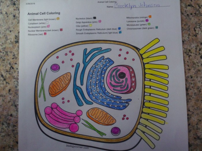
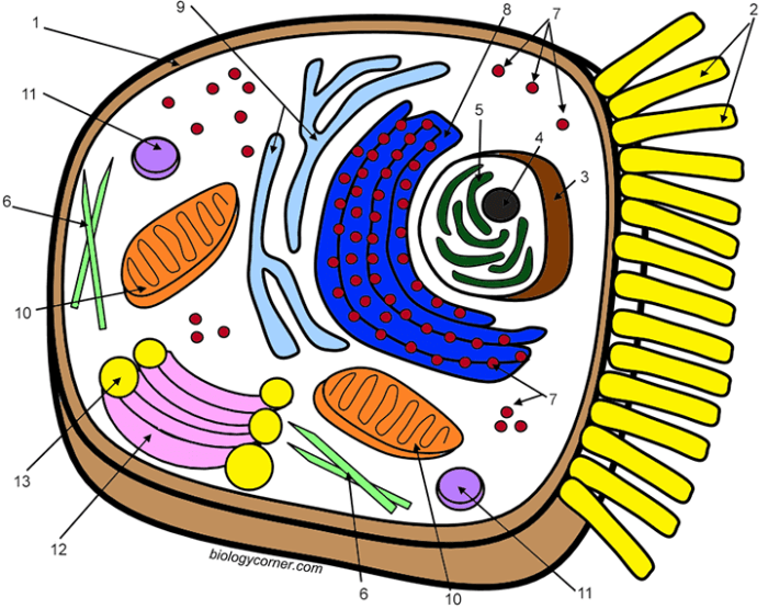
Animal celll coloring worksheet biologlycorner – Animal cells are the fundamental building blocks of animals, exhibiting a complex internal organization crucial for their diverse functions. Understanding their structure is key to grasping the intricacies of animal biology. This section will explore the major organelles and their roles within the animal cell, highlighting key differences compared to plant cells.
Animal cells, unlike plant cells, lack a rigid cell wall and a large central vacuole. This contributes to their greater flexibility and diverse shapes. However, they share many organelles with plant cells, each performing specialized tasks to maintain cell life.
Major Organelles and Their Functions
Animal cells contain various organelles, each with a specific function vital for cell survival and operation. These organelles work in a coordinated manner to ensure the cell’s overall health and function.
| Organelle | Function | Description | Analogy |
|---|---|---|---|
| Cell Membrane | Regulates the passage of substances into and out of the cell. | A selectively permeable barrier composed of a phospholipid bilayer. | A bouncer at a nightclub, selectively allowing entry. |
| Nucleus | Contains the cell’s genetic material (DNA) and controls cell activities. | The control center of the cell, housing chromosomes. | The mayor’s office of a city, directing all operations. |
| Cytoplasm | The gel-like substance filling the cell, containing organelles. | Aqueous solution holding organelles and facilitating transport. | The city itself, providing space and infrastructure. |
| Mitochondria | Generate energy (ATP) through cellular respiration. | The “powerhouses” of the cell, converting nutrients to energy. | Power plants of a city, providing electricity. |
| Ribosomes | Synthesize proteins. | Sites of protein production, found free in the cytoplasm or attached to the ER. | Construction workers building proteins within the city. |
| Endoplasmic Reticulum (ER) | Synthesizes and transports proteins and lipids. | A network of membranes involved in protein and lipid synthesis and transport. | A highway system transporting goods throughout the city. |
| Golgi Apparatus | Processes, packages, and distributes proteins and lipids. | Modifies and sorts proteins and lipids for secretion or use within the cell. | A post office sorting and distributing packages. |
| Lysosomes | Break down waste materials and cellular debris. | Contain enzymes that digest cellular waste and foreign materials. | Recycling center of the city, breaking down waste. |
Differences Between Plant and Animal Cells
Plant and animal cells share some similarities, but key differences exist due to their distinct roles and functions. These differences are mainly related to structural components and metabolic processes.
The most prominent difference is the presence of a rigid cell wall in plant cells, providing structural support and protection. Animal cells lack this cell wall, contributing to their flexible shapes. Plant cells typically possess a large central vacuole for water storage and maintaining turgor pressure, a feature absent in animal cells. Chloroplasts, responsible for photosynthesis, are present in plant cells but absent in animal cells, reflecting the difference in their energy acquisition strategies.
Finally, plant cells may contain plastids such as chromoplasts (pigment storage) and leucoplasts (starch storage), which are generally not found in animal cells.
Worksheet Design and Functionality
This section details the design and functionality of a coloring worksheet aimed at reinforcing learning about animal cell structures. The worksheet employs a simplified diagram to make the learning process engaging and accessible for students. The design prioritizes clarity and ease of use, ensuring that students can easily identify and color the different organelles.The coloring worksheet features a simplified diagram of an animal cell, large enough to accommodate coloring without overcrowding.
The intricate details of the animal cell coloring worksheet from BiologyCorner felt strangely familiar, a comforting routine. Then, a burst of vibrant color caught my eye – I remembered the playful energy of printable anime coloring sheets , a stark contrast yet somehow equally engaging. Returning to the scientific precision of the cell worksheet, I found a renewed appreciation for the beauty in both the microscopic and the fantastical.
The design is intended to be visually appealing and engaging for students of various ages and learning styles. The use of color helps students associate specific functions with particular organelles.
Organelles for Coloring and Color Key
The worksheet will include a simplified animal cell diagram with the following organelles clearly labeled and ready for coloring: nucleus, cytoplasm, cell membrane, mitochondria, ribosomes, endoplasmic reticulum (both rough and smooth), Golgi apparatus, lysosomes, and vacuoles. This selection represents a balance between providing sufficient detail for learning and avoiding overwhelming complexity.
- Nucleus: Purple
- Cytoplasm: Light Yellow
- Cell Membrane: Dark Blue
- Mitochondria: Red
- Ribosomes: Dark Green
- Rough Endoplasmic Reticulum: Light Green
- Smooth Endoplasmic Reticulum: Light Orange
- Golgi Apparatus: Brown
- Lysosomes: Dark Purple
- Vacuoles: Light Pink
This color key ensures consistency and facilitates accurate identification of organelles during the coloring process. The colors are chosen for their visual distinctiveness and to avoid potential confusion.
Educational Objectives and Learning Outcomes
This coloring worksheet aims to achieve several key learning outcomes. The primary objective is to enhance students’ understanding of animal cell structure and the functions of major organelles. The visual nature of the worksheet aids in memorization and retention of information.The worksheet facilitates active learning by engaging students in a hands-on activity. By coloring and labeling the organelles, students actively participate in the learning process, leading to improved comprehension and recall.
This method is particularly effective for visual learners. Successful completion of the worksheet demonstrates the student’s ability to:
- Identify and name major animal cell organelles.
- Associate specific functions with particular organelles.
- Develop a visual understanding of the relative sizes and positions of organelles within the cell.
- Improve memorization and retention of key biological concepts.
The worksheet’s design supports these objectives by providing a clear, simplified diagram and a corresponding color key, making it accessible and effective for a wide range of learners.
Activity Suggestions and Extensions: Animal Celll Coloring Worksheet Biologlycorner
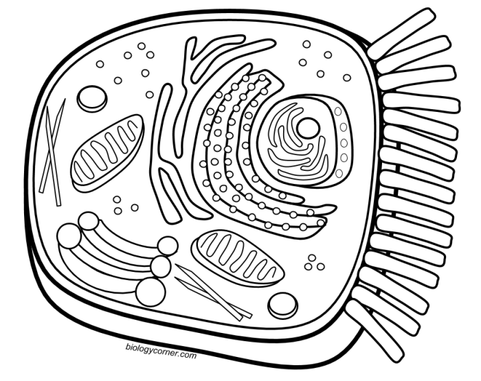
This animal cell coloring worksheet offers versatile applications within a biology classroom setting, extending beyond a simple coloring exercise to promote deeper understanding and engagement with cell structures. The worksheet can be adapted to suit various learning styles and assessment needs.The worksheet’s design allows for multiple uses, facilitating differentiated instruction and catering to diverse learning preferences. The visual nature of the activity helps students visualize complex biological structures, enhancing their comprehension and retention.
Classroom Activity Applications
This worksheet can be used in three distinct ways within a biology classroom. First, it can serve as an introductory activity, providing a visual foundation for subsequent lectures or discussions on animal cell structure and function. Second, it can be employed as a review activity, reinforcing previously learned concepts and identifying areas where students may need further clarification. Finally, it can function as a formative assessment tool, allowing teachers to quickly gauge student understanding of key cell components before moving on to more advanced topics.
3D Animal Cell Model Extension
To extend the learning experience, students can create a three-dimensional model of an animal cell using their completed worksheet as a guide. This activity encourages creativity and hands-on learning. Students could use various materials like clay, construction paper, or even recycled materials to represent the different organelles. The process of building the 3D model reinforces the spatial relationships between organelles and encourages collaborative learning if students work in groups.
The completed models can then be displayed in the classroom, serving as a visual representation of the collective learning. For example, the nucleus could be represented by a larger ball of clay, the mitochondria by smaller, elongated shapes, and the cell membrane by a translucent plastic bag.
Animal Cell Structure Quiz, Animal celll coloring worksheet biologlycorner
The following short quiz assesses student comprehension of animal cell structures following completion of the worksheet.
| Question | Answer |
|---|---|
| What is the control center of the animal cell? | Nucleus |
| Which organelle is responsible for energy production? | Mitochondria |
| What is the function of the cell membrane? | To regulate the passage of substances into and out of the cell. |
| Name two organelles involved in protein synthesis. | Ribosomes and Rough Endoplasmic Reticulum |
| What is the jelly-like substance that fills the cell? | Cytoplasm |
Illustrative Examples and Descriptions
This section provides detailed descriptions of key animal cell organelles, focusing on their structure and function. Understanding these components is crucial to grasping the overall operation of the cell.
The Nucleus: Cell Control Center
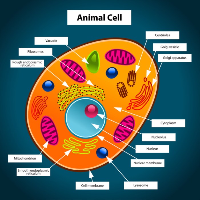
The nucleus is the control center of the animal cell, housing the cell’s genetic material, DNA. It’s a large, membrane-bound organelle typically located near the center of the cell. The nuclear envelope, a double membrane, surrounds the nucleus, regulating the passage of molecules between the nucleus and the cytoplasm. Within the nucleus, DNA is organized into chromosomes, which contain the instructions for building and maintaining the cell.
The nucleolus, a dense region within the nucleus, is responsible for ribosome biogenesis—the production of ribosomes, essential for protein synthesis. The nucleus directly controls gene expression, dictating which proteins are produced and when, ultimately governing all cellular activities. This control is achieved through the regulation of transcription, the process of copying DNA into RNA, which then serves as a template for protein synthesis.
Damage to the nucleus can severely impair or halt cellular function, leading to cell death.
Mitochondria: Powerhouses of the Cell
Mitochondria are often referred to as the “powerhouses” of the cell because they are responsible for generating most of the cell’s supply of adenosine triphosphate (ATP), the primary energy currency. These bean-shaped organelles are enclosed by a double membrane: an outer membrane and an inner membrane folded into cristae, which significantly increase the surface area for cellular respiration. Cellular respiration is a series of metabolic processes that convert nutrients, such as glucose, into ATP.
This process involves three main stages: glycolysis (in the cytoplasm), the Krebs cycle (in the mitochondrial matrix), and oxidative phosphorylation (on the cristae). Oxidative phosphorylation utilizes oxygen to generate a large amount of ATP. The efficiency of mitochondria is vital for cellular processes requiring significant energy, such as muscle contraction, nerve impulse transmission, and protein synthesis. Mitochondrial dysfunction can lead to a range of diseases, including mitochondrial myopathies and metabolic disorders.
Endoplasmic Reticulum and Golgi Apparatus: Protein Synthesis and Transport
The endoplasmic reticulum (ER) and Golgi apparatus work together in a coordinated manner to synthesize, modify, and transport proteins. The ER is a network of interconnected membranous sacs and tubules extending throughout the cytoplasm. There are two types of ER: rough ER and smooth ER. Rough ER is studded with ribosomes, which are the sites of protein synthesis. Proteins synthesized on the rough ER are often destined for secretion from the cell or for incorporation into membranes.
The smooth ER, lacking ribosomes, plays a role in lipid synthesis and detoxification. The Golgi apparatus, also known as the Golgi complex, is a stack of flattened, membrane-bound sacs called cisternae. Proteins synthesized on the rough ER are transported to the Golgi apparatus for further processing and modification. The Golgi apparatus adds sugars to proteins (glycosylation), sorts proteins, and packages them into vesicles for transport to their final destinations, such as the cell membrane or lysosomes.
The ER provides the initial protein synthesis, while the Golgi apparatus acts as a processing and packaging center, ensuring proteins reach their correct locations within or outside the cell. Disruptions in the ER-Golgi pathway can result in the accumulation of misfolded proteins and cellular dysfunction.
Pedagogical Considerations
Coloring worksheets offer a unique pedagogical approach to learning complex biological structures like the animal cell. This method leverages visual learning and kinesthetic engagement to enhance understanding and retention, particularly beneficial for students who respond well to hands-on activities and visual aids. The act of coloring encourages active participation, transforming passive reception of information into a more interactive learning experience.The effectiveness of coloring worksheets, however, can be compared and contrasted with other teaching methodologies.
While diagrams provide a static visual representation, coloring worksheets allow students to actively construct their understanding. Models offer a three-dimensional perspective, but can be expensive and less accessible. Videos, though dynamic, may lack the individual engagement offered by a hands-on activity. Each method caters to different learning preferences, and a multimodal approach incorporating several of these techniques would likely be most effective.
Comparison of Teaching Methods for Animal Cell Structure
The table below summarizes the advantages and disadvantages of various teaching methods used to illustrate animal cell structure. It highlights the strengths of the coloring worksheet approach in comparison to others, while acknowledging the limitations of relying solely on this method.
| Teaching Method | Advantages | Disadvantages |
|---|---|---|
| Coloring Worksheet | Engaging, promotes active learning, reinforces visual memory, adaptable to diverse learning styles, relatively inexpensive. | May lack depth of detail compared to models or videos, potential for inaccuracies if not carefully designed. |
| Diagrams | Provides a clear, concise visual representation, readily available in textbooks and online resources. | Can be static and less engaging, may not cater to visual learners effectively. |
| Models (3D) | Offers a realistic three-dimensional representation, enhances spatial understanding. | Can be expensive, may be fragile, less accessible to all students. |
| Videos/Animations | Dynamic and engaging, can illustrate complex processes effectively. | Requires technology, may be distracting, less opportunity for active learning. |
Adaptations for Diverse Learning Styles
The effectiveness of the coloring worksheet can be significantly enhanced by adapting it to suit various learning styles. Consider these adaptations to maximize inclusivity and engagement.The worksheet could be adapted to incorporate different levels of challenge, catering to both students who need extra support and those who are ready for more complex tasks. For instance, a simplified version with fewer labeled structures could be provided for students who require more support, while a more challenging version could include additional structures or require students to research and add information themselves.
Furthermore, different versions could be offered, some with pre-printed labels and others without, allowing students to work at their own pace and skill level. Finally, offering the worksheet in various formats (digital, printable) enhances accessibility.



0